What is anterior segment photography
Anterior segment photography is a procedure where the anterior chamber (front of the eye) is photographed to record the ocular structures of the eye and surrounding structures.
Where will I have the photographs taken?
Anterior segment photography is performed in the Clinical Photography Department. Depending on the requirements of the examination, your pupils may be dilated using eye drops, or imaging may be undertaken without dilation. This enables the clinical photographer to obtain the highest-quality images possible for assessment by the referring clinician.
How does this type of imaging differ from fundal photography and OCT?
Fundus imaging is used to photograph the back of the eye, including the retina and optic nerve, whereas anterior segment photography focuses on the front structures of the eye, such as the cornea, iris, lens, and surrounding tissues.
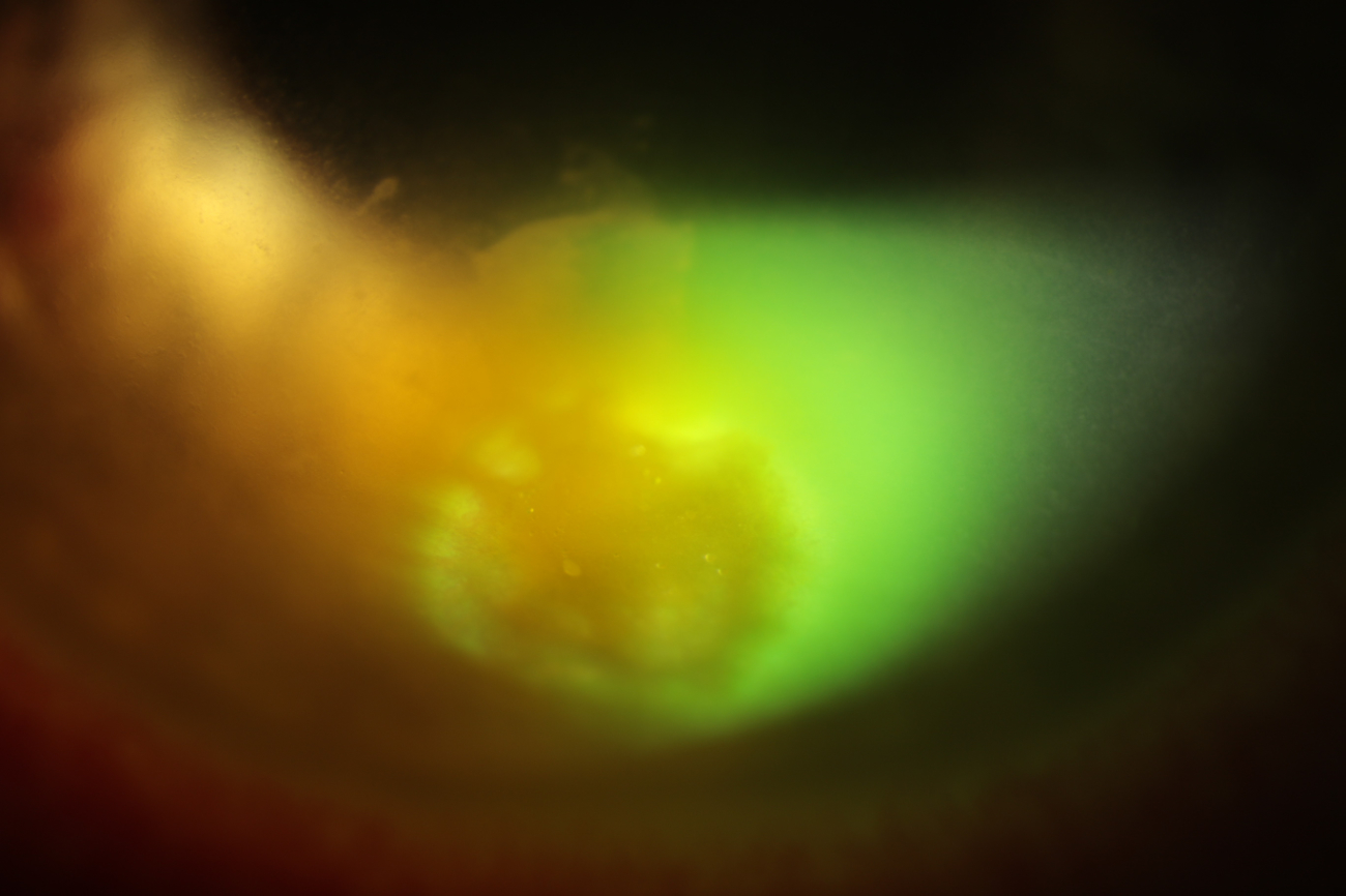
 Although anterior segment photography is primarily used to document the front of the eye, the retinal red reflex can sometimes be utilised to demonstrate certain ocular conditions. However, this technique does not provide detailed imaging of the retina itself.
In some cases, patients may have areas of iris thinning or defects. When clinically indicated, a technique known as transillumination may be performed. This involves directing light through the iris to highlight areas where the tissue is thin or absent, allowing these features to be documented photographically.
Anterior segment photography records the structures visible through the camera and slit-lamp optics. In contrast, Optical Coherence Tomography (OCT) uses near-infrared light to produce high-resolution cross-sectional images of ocular tissues. Similar in concept to ultrasound imaging, OCT uses light rather than sound waves and can provide detailed images of both anterior and posterior segment structures.
What does this type of imaging show?
This type of photography is used to image the anterior structures of the eye, including the sclera (the white part of the eye), cornea (the clear window at the front of the eye), iris (the coloured part of the eye that controls the amount of light entering the eye), eyelids, eyelashes, and surrounding tissues.
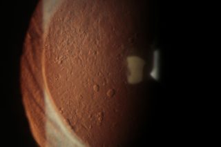
Anterior segment photography plays an important role in documenting and monitoring a wide range of eye conditions. For example, in patients with Fuchs’ endothelial corneal dystrophy, fluid can accumulate within the cornea, causing it to become cloudy and resulting in blurred vision and increased sensitivity to light. In more advanced stages of the condition, small blisters (bullae) may develop on the surface of the cornea. Anterior segment photographs can be used to record these changes and monitor disease progression over time.
Fuchs Dystrophy
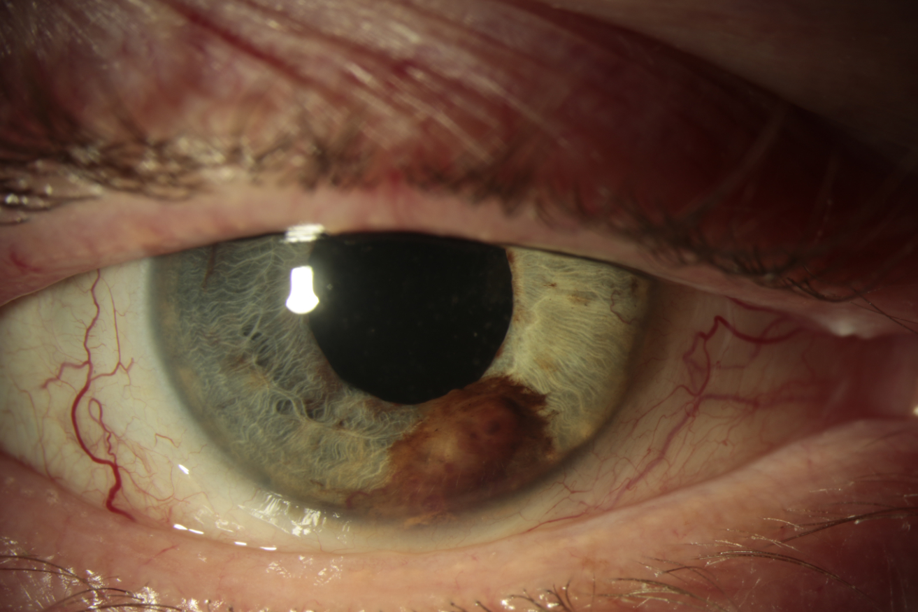
Tumour on Iris
Certain eye tumours, including melanomas arising from the iris or other anterior segment structures, can also be documented using anterior segment photography. The images provide a valuable record of the size, shape, colour, and appearance of the lesion, allowing clinicians to monitor for any changes.
Anterior segment photography is also used to assess a patient’s response to treatment. This includes monitoring the resolution of serious eye infections, evaluating healing following surgery, and documenting the adaptation, health, and potential rejection of corneal grafts. By providing a detailed visual record, anterior segment photography supports diagnosis, treatment planning, and ongoing clinical management.
Finally, in cases of trauma to the eye, anterior segment photography is useful to document damage to the eye before and after surgical consultations.
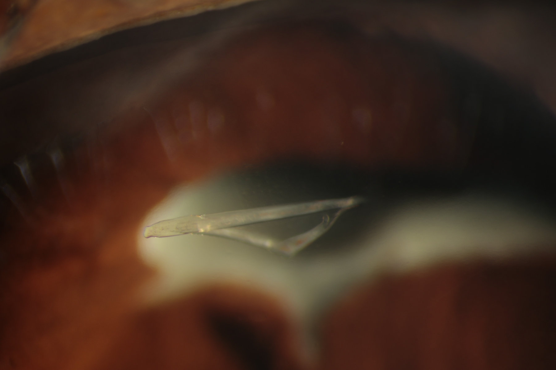
Glass Fragment
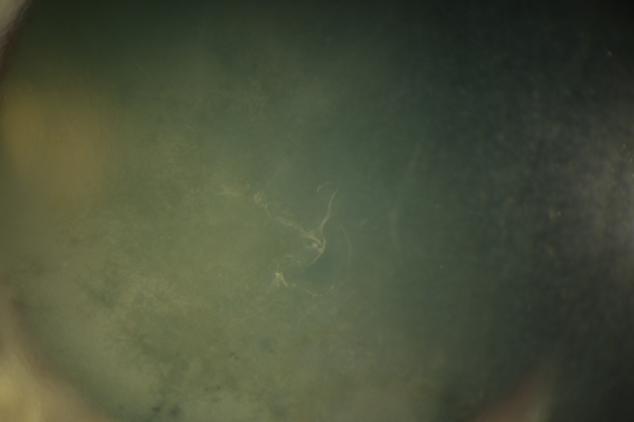
Eye infection
Are there any risks associated with this type of photography?
There is no risk associated with this form of imaging; however, patients with light sensitivity may experience some discomfort.