
What is Indocyanine Green Angiography (ICG)?
Indocyanine Green Angiography is the process where a contrast dye is used to image the choroid to identify any abnormalities that may require intervention and treatment to save a patient’s sight.
This diagnostic procedure utilizes a specialized fundus camera or scanning laser ophthalmoscope to capture rapid-sequence photographs of the choroid following an intravenous injection of Verdye Indocyanine Green dye.
Photographic or video images taken as the dye courses through the eye can demonstrate abnormalities within the choroid.
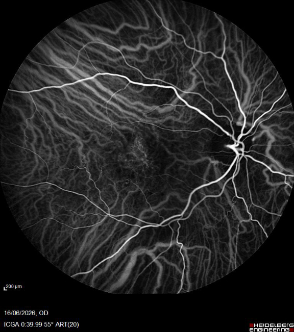
Fuchs Dystrophy
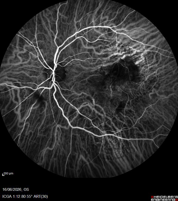
Age Related Macular Degeneration ICG LE
Where will I have the photographs taken?
Indocyanine Green Angiography (ICG) is carried out in the Clinical Photography Department and in the Macular Treatment Centre. Your pupils will be dilated (widened) using eye drops to allow clinical photographers to obtain the clearest possible images for the requesting doctor.
How does this test differ from fluorescein angiography photography?
Indocyanine Green Angiography (ICG) is a diagnostic procedure used to examine the choroidal circulation at the back of the eye. It’s also used to identify areas that may require treatment for specific conditions that affect the retina. It involves injecting an infra-red contrast dye called Indocyanine Green into the bloodstream.
A nurse or doctor will insert a small intravenous cannula, usually into a vein in your arm or hand, to administer the dye. Once injected, the dye travels through the blood vessels to the eye. You will then be asked to look into a specialised camera while a series of photographs are taken.
The principle is similar to fluorescein angiography, but here we are interested in imaging the structures of the choroid. This is the outer vascular layer of the eye located between the sclera and retina; this layer provides oxygen and nutrients to the outer layers of the retina.
These images capture the movement of the dye through the choroidal blood vessels, helping clinicians assess and diagnose various eye conditions.
What does Indocyanine Green (ICG) show?
Indocyanine Green Angiography (ICG) helps us image the choroid and it’s used to assess conditions including Age Related Macular Degeneration (AMD), Polypoidal Choroidal Vasculopathy (PCV), inflammatory conditions including Birdshot Chorioretinopathy, and it’s also widely used in Ocular Oncology to help identify choroidal hemangiomas and differentiate them from simulating tumours such as choroidal melanoma or metastasis.
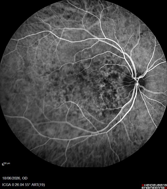
Choroidal Hemangioma Optic Nerve
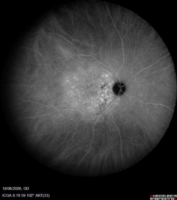
Choroidal Hemangioma Widefield
Are there any risks associated with Indocyanine Green Angiography?
Yes, like any other contrast injection there are risks associated with ICG angiography. Your doctor will advise you of these risks and possible side effects of the test, and you will also be required to sign a consent form before the test is carried out.
Why do you use different cameras?
Like fundal photography, the type of retinal camera used for Indocyanine Green depends on the patient’s suspected or confirmed diagnosis. Different eye conditions affect different areas of the retina, so selecting the most appropriate imaging system helps clinicians obtain the most useful information.
For example, in patients with age-related macular degeneration (AMD), imaging is usually focused on the central choroid beneath the retina, as this is the area affected by the condition. A camera with a narrower field of view is often sufficient to document changes in this region.
Indocyanine Green Angiography can generally be divided into two main areas:
Central pole imaging focuses on the central choroid, primarily documenting the structures beneath the optic nerve, macula, and fovea. This type of imaging is commonly used to assess conditions affecting central vision.
Peripheral retinal imaging uses ultra-widefield retinal cameras to capture images of the outer areas of the choroid. These systems allow clinicians to visualise and document the peripheral choroid, which can be important for detecting and monitoring conditions that may not be visible in standard central images.