This website uses cookies so that we can provide you with the best user experience possible. Cookie information is stored in your browser and performs functions such as recognising you when you return to our website and helping our team to understand which sections of the website you find most interesting and useful.
What is an OCT scan?
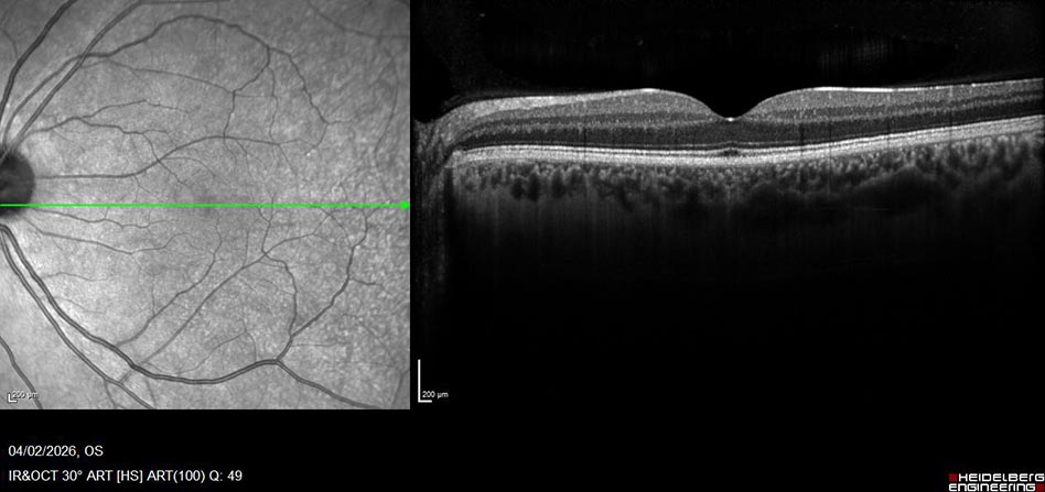
Optical Coherence Tomography, or OCT for short is a scan of the retina at the back of your eye. OCT scans can be taken of both the anterior chamber (front) and posterior chamber (back) of your eye to diagnose and monitor diseases that may affect patients eyes. OCT uses coherent interferometry to construct a cross-sectional view of the ocular structures. Its quick and non invasive with scans being captured in-vivo. Below is an example of a normal OCT scan on patient with no retinal pathology. The scan is focused on the fovea and macula, the fovea responsible for our central vision.
Where will I have the scans taken?
OCT scans are carried out in eye out patients and Macular Treatment Centre. Your eyes will be dilated (pupils widened) to allow our clinical photographers to get the best scans possible for the requesting doctors. In certain instances, if your eye pressure is of concern, the requesting doctor may as for the OCT to be taken without dilating drops. In this case the clinical photographers will do their best to get a good quality scan on your retina.
Learn more about the structure of the retina. (Link to American Academy of Ophthalmology)
What does the OCT look for?
OCT scan be broken down into two areas of scanning:
- Retinal scanning of the back of the eye. (Retina & Optic Nerve)
- Anterior scanning of the front of the eye. (Cornea, Iris, Cilary Body, Sclera & Lens)
For retinal patients, that have conditions that affect the back of their eye, OCT is useful to monitor patients response to treatment, document and changes in the structures of the retina. A large cohorts of patients that attend the Royal Victoria Eye & Ear would be those diagnosed with Age Related Macular Degeneration (AMD), an age related condition that affects the patients central vision.
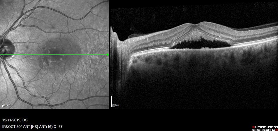
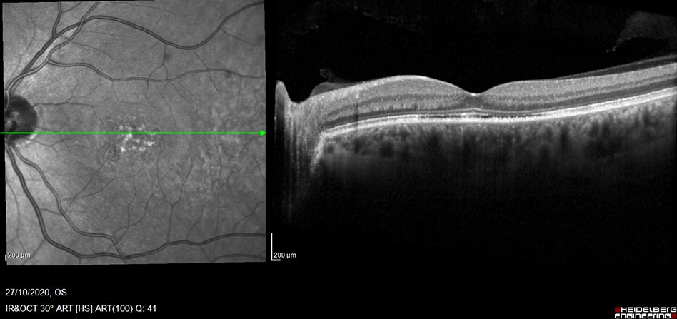
There are two types of AMD, a ‘wet’ or neovascular form identified by the presence of fluid in the retina and a ‘dry’ form which presents no fluid. Treatments vary for each specific retinal condition, these could include intravitreal into the eye, drops administered topically, IV infusions of laser to the retina. All of these treatments can be monitored with the use of OCT scanning.
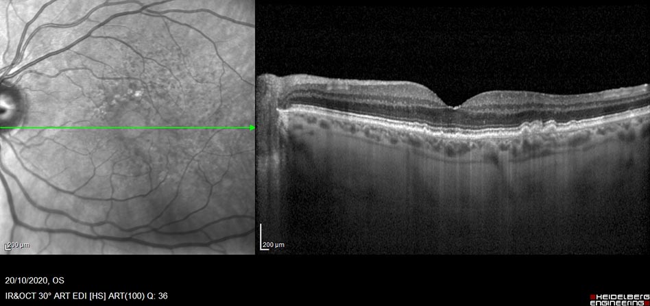
Dry AMD OCT
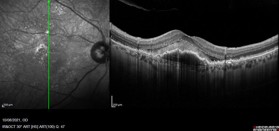
WET OCT
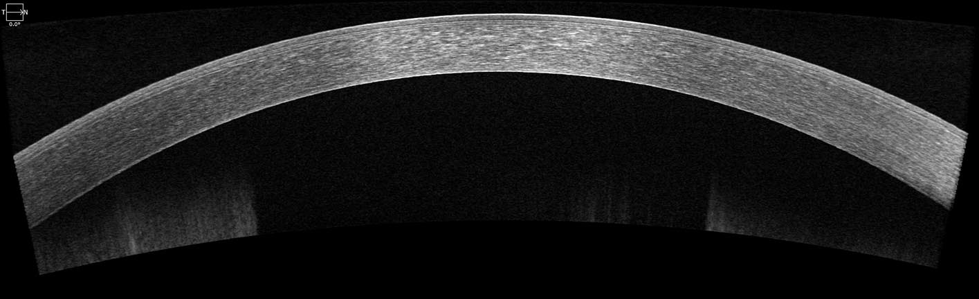
Another useful application is the use of OCT to scan the cornea, this is the clear structure at the front of the eye. There are five layers in the cornea: epithelium, bowman’s layer, stroma, descents membrane and endothelium. These scans are particularly useful in patients who have had corneal transplant surgery, the surgeon can assess outcome of the surgery by viewing the endothelium, checking for detachments.
Finally OCT used in the education of patients about their diagnosis. In some instances it may be difficult for patients to comprehend and visualize what that are having treatment for. Healthcare professionals can use OCT to illustrate patients response to treatment and current state of the retina.
OCT scan pre treatment
OCT Post treatment
Useful external links:
https://www.opsweb.org/page/octimaging
https://www.aao.org/eye-health/anatomy/fovea
https://www.aao.org/eye-health/anatomy/parts-of-eye