This website uses cookies so that we can provide you with the best user experience possible. Cookie information is stored in your browser and performs functions such as recognising you when you return to our website and helping our team to understand which sections of the website you find most interesting and useful.
What is fundal photography?
Retinal photography is the process by which a specialised camera is used to capture detailed images of the blood vessels and structures at the back of the eye, known as the retina. These images help clinicians to assess eye health, monitor disease progression, and document changes over time.
There are several different types of retinal cameras used to image the retina, each designed for specific clinical purposes and levels of detail.
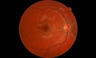 Right Retina |
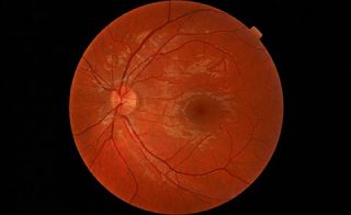 Left Retina |
Where will I have the photographs taken?
Retinal photography is carried out in the Clinical Photography Department and in the Macular Treatment Centre. In most cases, your pupils will be dilated (widened) using eye drops to allow clinical photographers to obtain the clearest possible images for the requesting doctor.
In certain circumstances, if there are concerns about your eye pressure, the requesting doctor may ask for photographs to be taken without dilating drops. When this happens, some retinal camera systems cannot be used, as they require pupil dilation to capture the necessary images. In these cases, alternative non-dilated imaging systems will be used where appropriate.
What does fundal photography show?
Retinal photography documents the back of the eye and shows the normal anatomical structures of the retina, including the veins, arteries, optic nerve head, macula, and fovea. These images provide clinicians with a detailed visual record of eye health.

If clinicians suspect that a patient may have complications related to a particular eye condition, retinal photography allows abnormal findings to be documented and monitored over time. This helps support diagnosis, treatment planning, and ongoing review.
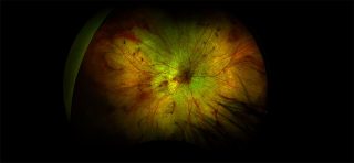
Different imaging filters can be used to highlight specific structures at the back of the eye. These include red-free imaging, which enhances the visibility of blood vessels and nerve
fibre layers, and autofluorescence imaging, which helps assess the health of the retinal pigment epithelium and detect certain retinal diseases.
Retinal photography can generally be divided into two main areas:
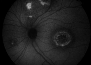
Central pole imaging focuses on the central retina, primarily documenting the optic nerve, macula, and fovea. This type of imaging is commonly used to assess conditions affecting central vision and optic nerve health.
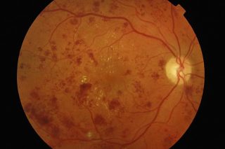 Colour Fundas |
 Red Free Photo |
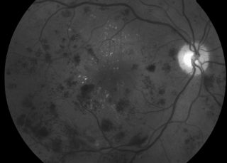
 Autoflourescence |
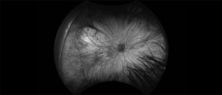
Peripheral retinal imaging uses ultra-widefield retinal cameras to capture images of the outer areas of the retina. These systems allow clinicians to visualise and document the peripheral retina, which can be important for detecting and monitoring conditions that may not be visible in standard central images.
 Colour Widefield Optos |
 AF Widefield Optos |
Why do you use different cameras?
The type of retinal camera used depends on the patient’s suspected or confirmed diagnosis. Different eye conditions affect different areas of the retina, so selecting the most appropriate imaging system helps clinicians obtain the most useful information.
For example, in patients with age-related macular degeneration (AMD), imaging is usually focused on the central retina (macula), as this is the area affected by the condition. A camera with a narrower field of view is often sufficient to document changes in this region.
In contrast, patients with diabetic retinopathy often require widefield or ultra-widefield imaging. This allows clinicians to examine the peripheral retina and check for signs such as peripheral ischaemia, new blood vessel growth, or other changes that may not be visible in standard central images.
Colour imaging also plays an important role in the assessment and treatment of certain eye conditions, including some ocular tumours. Accurate colour representation helps clinicians document lesions, monitor changes over time, and support diagnosis alongside other clinical investigations.
Useful external links: