This website uses cookies so that we can provide you with the best user experience possible. Cookie information is stored in your browser and performs functions such as recognising you when you return to our website and helping our team to understand which sections of the website you find most interesting and useful.
What is a fundus Fluorescein Angiography
What is Fundus Fluorescein Angiography?
Fundus Fluorescein Angiography is the process where a contrast dye is used to image the retina to identify any abnormalities that may require intervention and treatment to save a patient’s sight. Since its introduction in the early 1960s, fluorescein angiography has become an essential tool in the understanding, diagnosis and treatment of retinal disorders.
This diagnostic procedure utilises a specialised fundus camera or scanning laser ophthalmoscope to capture rapid-sequence photographs of the retina following an intravenous injection of fluorescein sodium.
Photographic or video images taken as the dye courses through the eye can demonstrate abnormalities within the neurosensory retina, pigment epithelium, sclera, choroid, and optic nerve, providing clinically useful information for nearly the entire spectrum of posterior segment disorders.
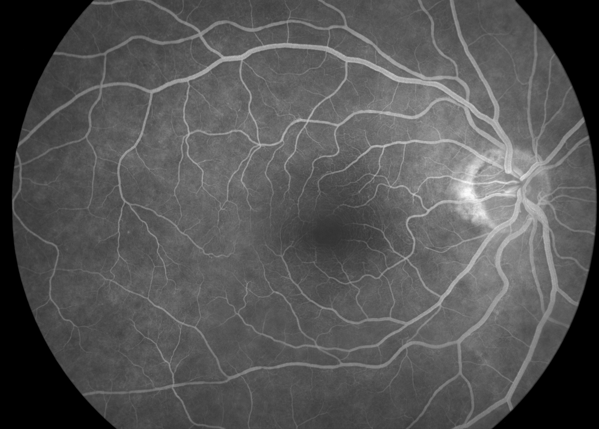
Fluorescein Angiography showing no leakage
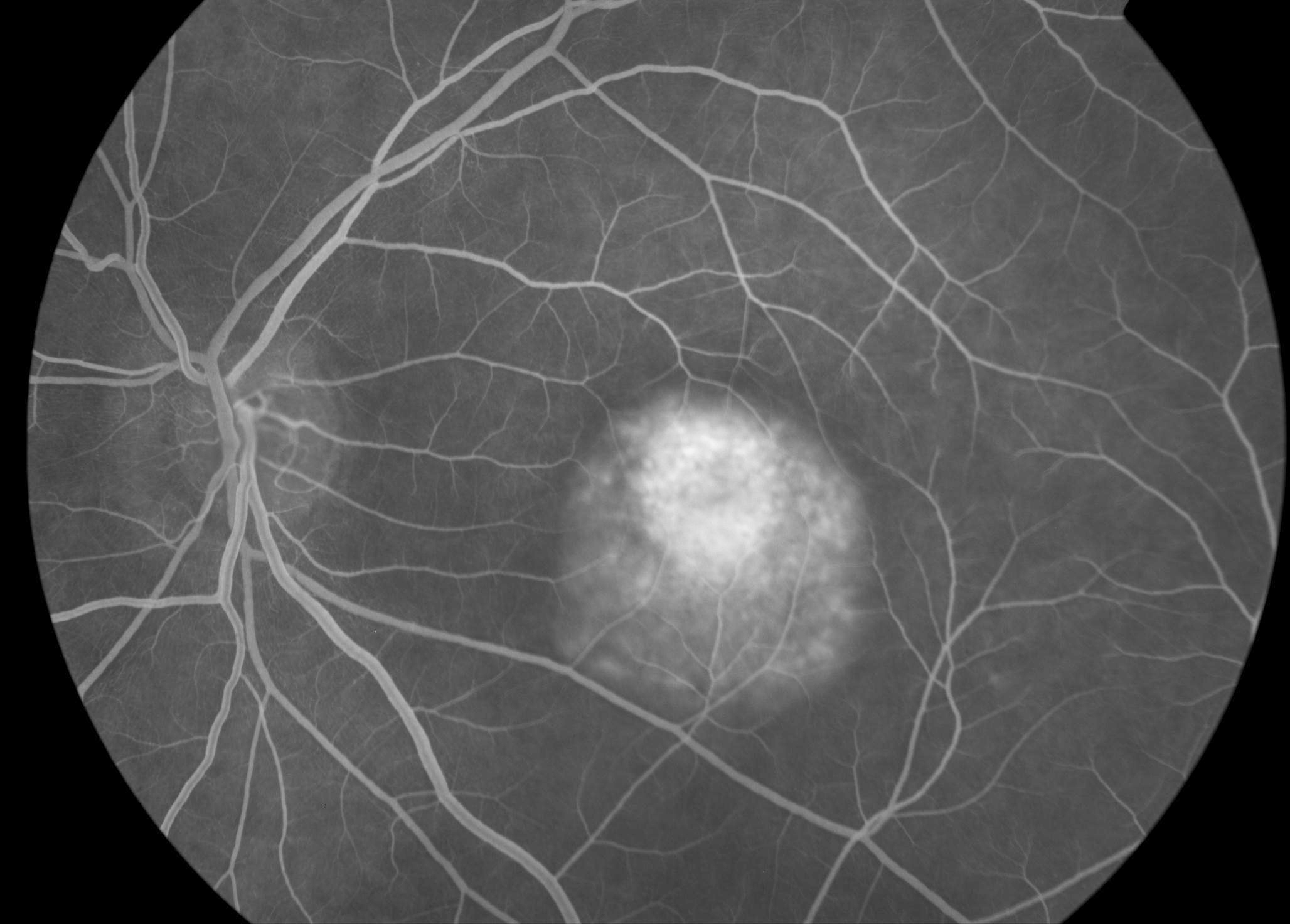
Fluorescein Angiography showing leakage at macula (Patients central vision)
Where will I have the photographs taken?
Fluorescein Angiography is carried out in the Clinical Photography Department and in the Macular Treatment Centre. Your pupils will be dilated (widened) using eye drops to allow clinical photographers to obtain the clearest possible images for the requesting doctor.
How does this test differ from fundal photography?
Fundus Fluorescein Angiography is a diagnostic procedure used to examine the retinal circulation at the back of the eye. It is also used to identify areas that may require treatment for specific conditions that affect the retina. It involves injecting a fluorescent contrast dye called fluorescein into the bloodstream.
A nurse or doctor will insert a small intravenous cannula, usually into a vein in your arm or hand, to administer the dye. Once injected, the dye travels through the blood vessels to the eye. You will then be asked to look into a specialised camera while a series of photographs are taken.
These images capture the movement of the dye through the retinal blood vessels, helping clinicians assess and diagnose various eye conditions. Learn more about the fluorescein dye.
What does Fluorescein Angiography show?
Fluorescein angiography is most commonly used to diagnose and assess retinal or choroidal vascular diseases such as diabetic retinopathy, age-related macular degeneration, hypertensive retinopathy and vascular occlusions. It is carried out in conjunction with OCT to determine the extent of damage to the retinal structures and choroid, to develop a treatment plan, monitor disease progression and patient response to treatment. Areas that become brighter (hyper-fluorescent) indicate abnormal leakage in the retina and may require treatment to stop further sight loss.
In diabetic retinopathy, the angiogram is useful in identifying the extent of ischaemia, the location of microaneurysms, the presence of neovascularisation (new abnormal blood vessels) and the extent of macular oedema. In age-related macular degeneration, angiography is useful in identifying the presence, location and characteristic features of choroidal neovascularisation for possible treatment with laser photocoagulation, photodynamic therapy, or antiangiogenic medications.
Some conditions Fluorescein Angiography is used to help diagnose and treat:
- Diabetic retinopathy
- Age-related macular degeneration
- Neovascular membranes (myopia, histoplasmosis)
- Central retinal vein occlusion
- Branch retinal vein occlusion
- Central serous retinopathy
- Branch retinal artery occlusion
- Retinal arterial macroaneurysms
- Choroidal tumours
- Chorioretinal inflammatory conditions
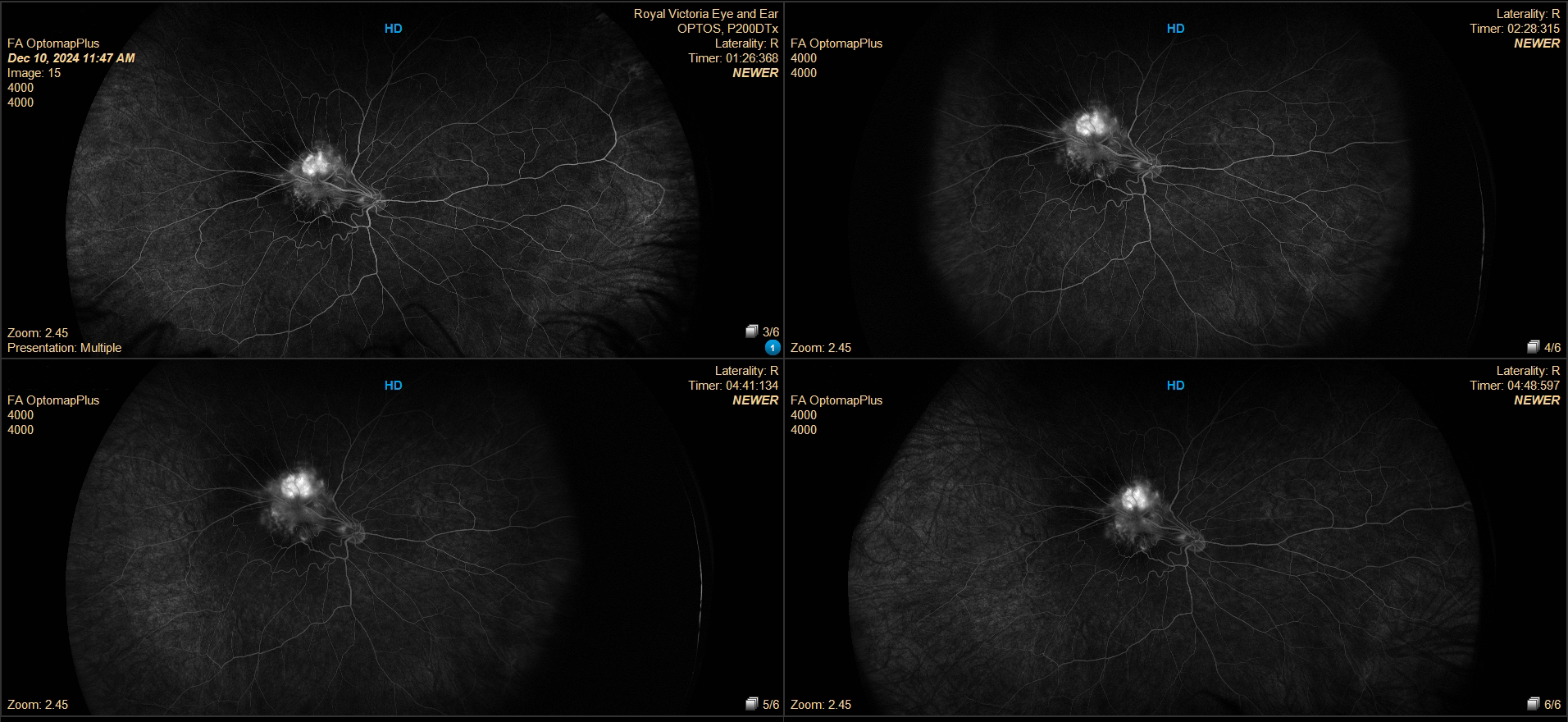
Four up FFA
Are there any risks associated with Fluorescein Angiography?
Yes, like any other contrast injection there are risks associated with Fluorescein Angiography. Your doctor will advise you of these risks and possible side effects before the test is carried out. You will also be required to sign a consent form.
Fluorescein Angiography imaging can generally be divided into two main areas:
Central pole imaging focuses on the central retina, primarily documenting the optic nerve, macula, and fovea. This type of imaging is commonly used to assess conditions affecting central vision and optic nerve health.
Peripheral retinal imaging uses ultra-widefield retinal cameras to capture images of the outer areas of the retina. These systems allow clinicians to visualise and document the peripheral retina, which can be important for detecting and monitoring conditions that may not be visible in standard central images.
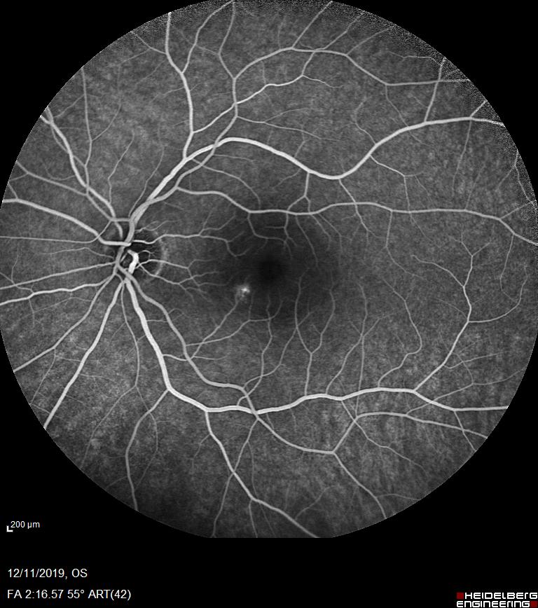
Fluorescein Angiogram showing focal leakage in CSR patient
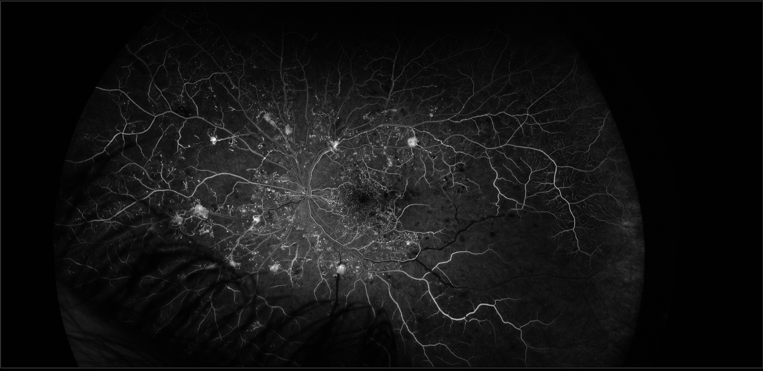
Widefield Fluorescein Angiogram of Diabetic Retina.
Why do you use different cameras?
Like fundal photography, the type of retinal camera used for Fluorescein Angiography depends on the patient’s suspected or confirmed diagnosis. Different eye conditions affect different areas of the retina, so selecting the most appropriate imaging system helps clinicians obtain the most useful information.
For example, in patients with age-related macular degeneration (AMD), imaging is usually focused on the central retina (macula), as this is the area affected by the condition. A camera with a narrower field of view is often sufficient to document changes in this region. Fluorescein Angiography can help confirm wet AMD, which requires immediate treatment to stop rapid vision loss.
In contrast, patients with diabetic retinopathy often require widefield or ultra-widefield imaging. This allows clinicians to examine the peripheral retina and check for signs such as peripheral ischaemia, new blood vessel growth, or other changes that may not be visible in standard central images. Fluorescein Angiography can also show leakage in the wider peripheral retina.